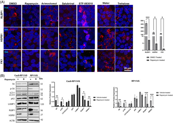
FIGURE 8.

Elimination of aggregates in RP11‐RPE cells through application of Rapamycin. Representative immunofluorescence images and quantification analysis of RP11VS‐RPE cells showing a significant decrease of cytoplasmic aggregates containing RLBP1 (red), HSPB1(red) and FK1 (red) upon daily treatment with Rapamycin (500 nM) for 7 days. No apparent differences were observed in cytoplasmic aggregates after seven days treatment with Arimoclomol (1 μM), Salubrinal (25 μM), STF‐083010 (50 μM) and trehalose (50 mM). DMSO was used as a vehicle control for Rapamycin, Arimoclomol, Salubrinal and STF‐083010, and distilled water was used as vehicle control for Trehalose. Cell nuclei were counterstained with Hoechst. Scale bars: 20 μm. Quantification of these results is shown on the right‐hand side graph presented as mean ± SEM (n = 3). Statistical significance was assessed using paired Student t‐test. *P < 0.05, ***P < 0.001. (B) Western blot and quantification analysis of 7 days Rapamycin‐treated isogenic control and RP11VS‐RPE cells showing the decrease in the expression of S6, p‐S6 RLBP1, HSPB1 and increase in LC3‐II expression in RP11VS‐RPE cells. Actin B was used as a loading control. Data represent the mean ± SEM (n = 3). Statistical significance was assessed using two‐way ANOVA. *P < 0.05, **P < 0.001, ***P < 0.001. Please refer to Figures S7 and S8 for the same analysis in two additional RP11‐RPE cells.
