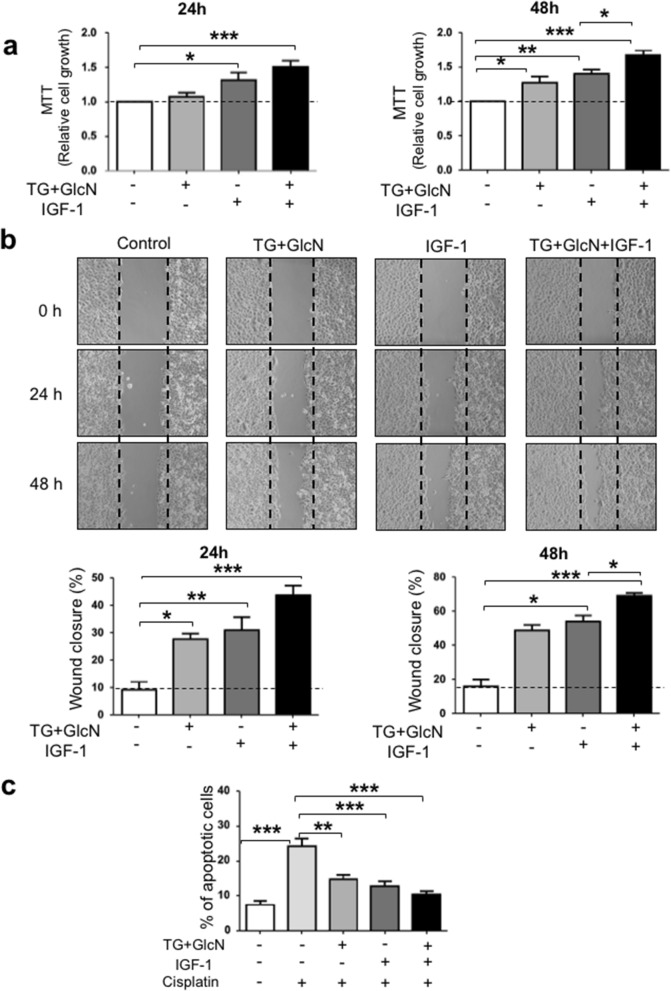
Figure 2.
O-GlcNAcylation-inducing treatments increase cell growth and migration and inhibit apoptosis in CaSki cells. (a) Cells were cultured in 1% FBS and cultured in the absence and presence of O-GlcNAcylation-inducing agents (TG 10 µM and GlcN 5 mM) and IGF-1 (5 nM). MTT assay was used to determine the cell growth at 24 h and 48 h. Results are the mean ± SEM of 5 independent experiments. (b) Wound healing assay was performed as described in “Methods”. Representative images of the wound at 0, 24 and 48 h are shown. Migration is expressed as the wound closure percentage at 24 h and 48 h. The results are presented as mean ± SEM of 4 independent experiments. (c) CaSki cells were treated for 24 h with O-GlcNAcylation-inducing agents, IGF-1 and Cisplatin, and then stained with Annexin V and propidium iodide for FACS analysis. The results are presented as mean ± SEM of 4 independent experiments. Statistical analysis was performed using ANOVA followed by Tukey’s post-test. *, **, ***p < 0.05, p < 0.01 and p < 0.001, respectively.

