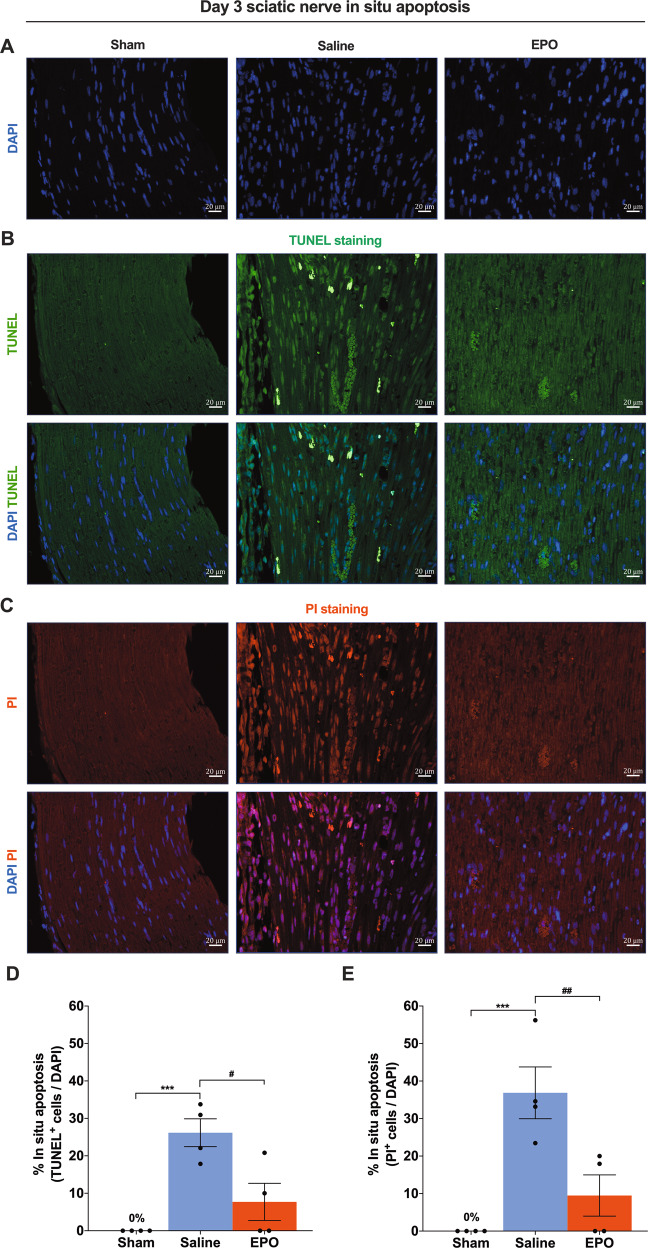
Fig. 1. On day 3, EPO effectively attenuates SN in situ apoptosis following SNCI.
A DAPI staining shows cellular infiltration of SN. B, C Representative IF images of TUNEL and PI staining of SN shows that EPO treatment (5000 IU/kg/IP, immediately after surgery and on post-surgery day 1 and 2) significantly attenuates apoptosis as compared to saline treatment (normal saline, 0.1 ml/IP/mouse) and the result of the percentage of in situ apoptosis (TUNEL and PI+/DAPI) are mentioned in the bar graph (D, E). SN of Sham animals shows zero percentage of apoptosis. Each image represents six images from four different SN, a total of 24 images/group. Scale bar, 20 μm; magnification, 40x. One-way ANOVA, Tukey’s multiple comparisons test. Data were expressed as means ± SEM, ***P < 0.0002 vs. sham, #P < 0.05 and ##P < 0.0021 vs. Saline, n = 4/group.

