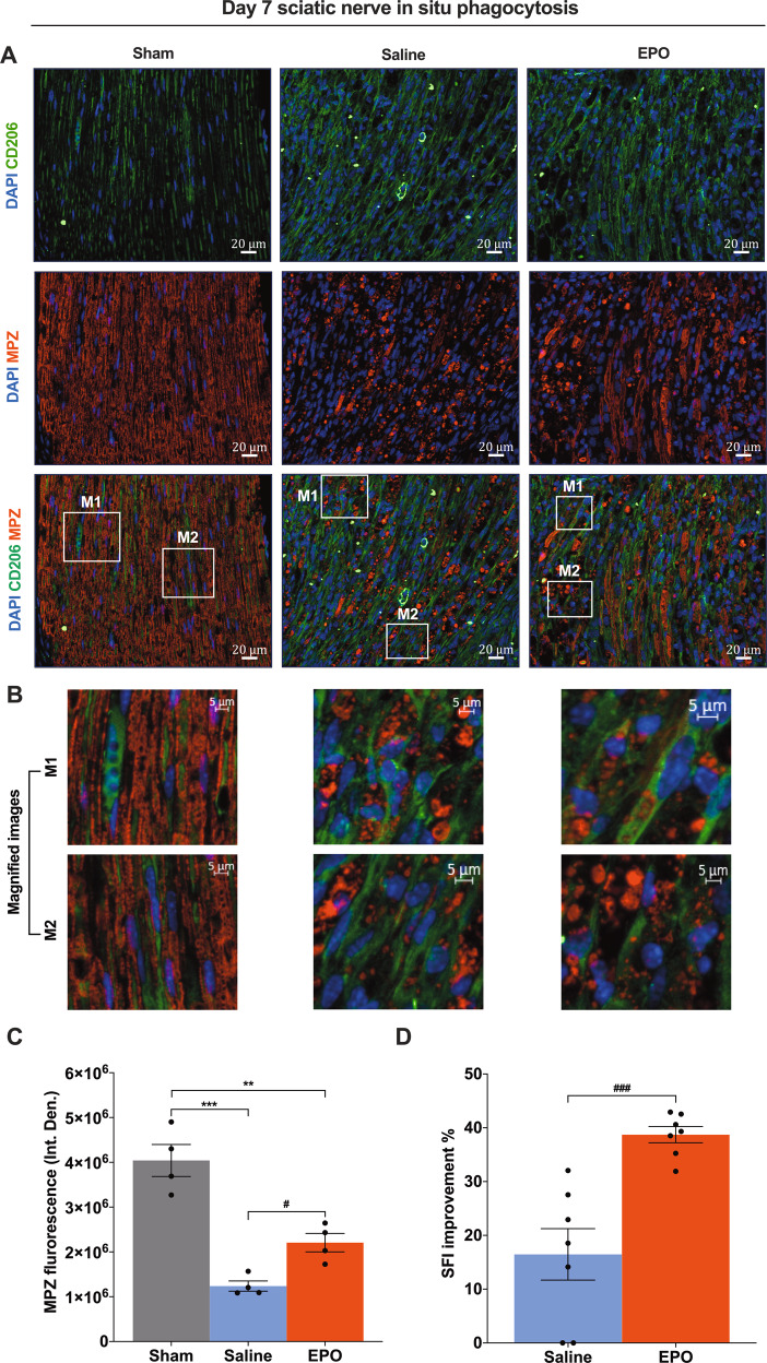
Fig. 4. On day 7, EPO significantly activates CD206+ macrophage phagocytosis of myelin debris, which improves myelination and functional recovery following SNCI.
A Representative IF images of CD206 (M2 phenotype macrophages) and MPZ (Myelin) staining of SN shows that EPO treatment (5000 IU/kg/IP, immediately after surgery and on post-surgery day 1 and 2) effectively control phagocytosis of myelin debris and which significantly improves myelination as compared to saline treatment (normal saline, 0.1 ml/IP/mouse) and are depicted in magnified images (B) and the results of the integrated density of MPZ are depicted in the bar graph (C). M1 and M2 are random subset images of the SN section. D EPO treatment significantly improves %SFI. Each image represents six images from four different SN, a total of 24 images/group. Scale bar, 20 μm; magnification, 40x. One-way ANOVA, Tukey’s multiple comparisons test. Data were expressed as means ± SEM, ***P < 0.0002, Saline vs. sham; **P < 0.0021, EPO vs. sham; #P < 0.05, EPO vs, saline, n = 4/group (CD206/MPZ staining analyses). Unpaired t-tests, ###P < 0.0002, EPO vs, saline, n = 7/group (SFI analyses).

