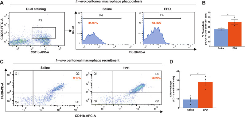
Fig. 8. EPO treatment augments M2 phenotype peritoneal macrophage phagocytosis, in vivo.
A Flow cytometry gating strategy of dual staining (CD11b + CD206+), where that shows (P4, histogram) EPO treatment (5000 IU/kg/IP, immediately after surgery and on post-surgery day 1 and 2) significantly augments percent phagocytosis of intraperitoneally injected apoptotic SNSCs (PKH26 labeled) as compared to saline treatment (normal saline, 0.1 ml/IP/mouse) and the results are denoted in the bar graph (B). C Flow cytometry data shows EPO significantly recruits PMØs (CD11b + F4/80+) following intraperitoneal injection of apoptotic SNSCs (PKH26 labeled) as compared to saline treatment and the results are denoted in the bar graph (D). Unpaired t-tests. Data were expressed as means ± SEM, #P < 0.05, Saline vs. EPO, n = 3/group.

