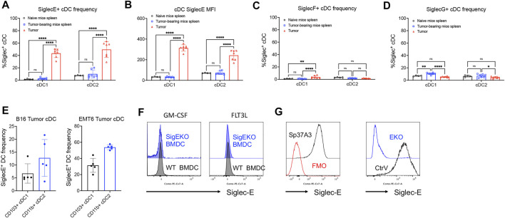
FIGURE 2.
Inhibitory Siglec-E expression is significant on mouse tumor-associated DCs. (A–D) The expression patterns of several murine inhibitory Siglecs on cDCs isolated from naive C57BL/6 mice spleens, MC38 tumor-bearing mouse spleens, and primary subcutaneous tumors were analyzed by flow cytometry. (A) SiglecE + cDC frequency, (B) SiglecE MFI, (C) SiglecF + cDC frequency, and (D) SiglecG + cDC frequency. (E) Siglec-E expression on tumor cDC subsets from B16 melanoma and EMT6 breast cancer mouse models. (F) Siglec-E expression of BMDCs from wildtype (WT, black line) and systemic Siglec-E knockout (EKO, blue line) mice after 7-day in vitro culture supplemented with GM-CSF or FLT3L. (G) The expression of Siglec-E on Sp37A3 cell line (black line) versus FMO control (red line). Siglec-E expression of Siglec-E knockout (EKO) Sp37A3 cells and empty control vector (CtrV)-transduced Sp37A3 cells. Data are presented as mean ± SD, and two-way ANOVA was used for two-way comparisons (*p < 0.0332, **p < 0.0021, ***p < 0.0002, and ****p < 0.0001).

