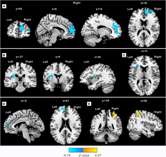
FIGURE 1.
Practitioners demonstrate a significantly different amplitude of low-frequency fluctuations (ALFF) after an 8-week practice. The clusters detected with decreased ALFF include: (A) right anterior cingulate gyrus (ACC.R), extending into the right middle frontal gyrus; (B) left posterior insula (pIC.L), extending into the left putamen; (C) left anterior insula (aIC.L), extending ventrolateral into the left middle frontal gyrus; (D) peak in the left superior medial frontal gyrus (SFGmed.L), extending into the left superior dorsolateral frontal gyrus. A cluster located in the right postcentral gyrus (PostCG.R) extending into the right superior parietal gyrus (E) showed increased ALFF. Sections are shown in sagittal, axial, and coronal planes with Montreal Neurological Institute (MNI) coordinates of the selected sections representing the peak in the x-, y-, and z- direction.

