
FIGURE 4.
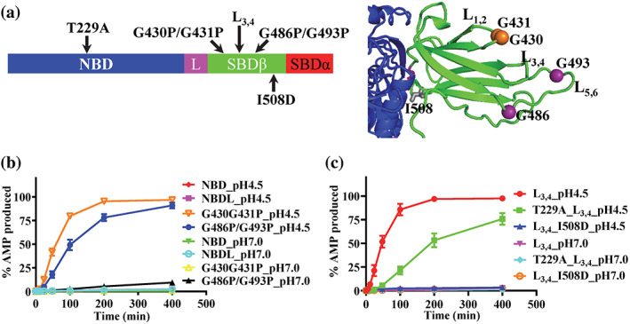
The nucleotide‐binding domain–substrate‐binding domain (NBD–SBD) contacts are required for the ATP to AMP hydrolysis by Binding immunoglobin protein (BiP). (a) The locations of the BiP mutations. Left, the BiP mutations were labeled in the domain organization of BiP. The domain coloring and labeling were the same as Figure 1a. Right, ribbon diagram of the BiP‐AMP structure. Only the SBDβ (green) and the part of the NBD (blue) forming contacts with SBDβ were shown. (b) The ATP hydrolysis to AMP was not observed without SBD but enhanced by the G430P/G431P and G486P/G493P mutations in the peptide‐binding site. (c) The T229A and I508D mutations compromised the ATP to AMP hydrolysis by BiP. For both (b) and (c), the AMP production was plotted over time at pH 4.5 and pH 7.0. Mean ± SEM from three independent experiments with more than two different protein purifications were used