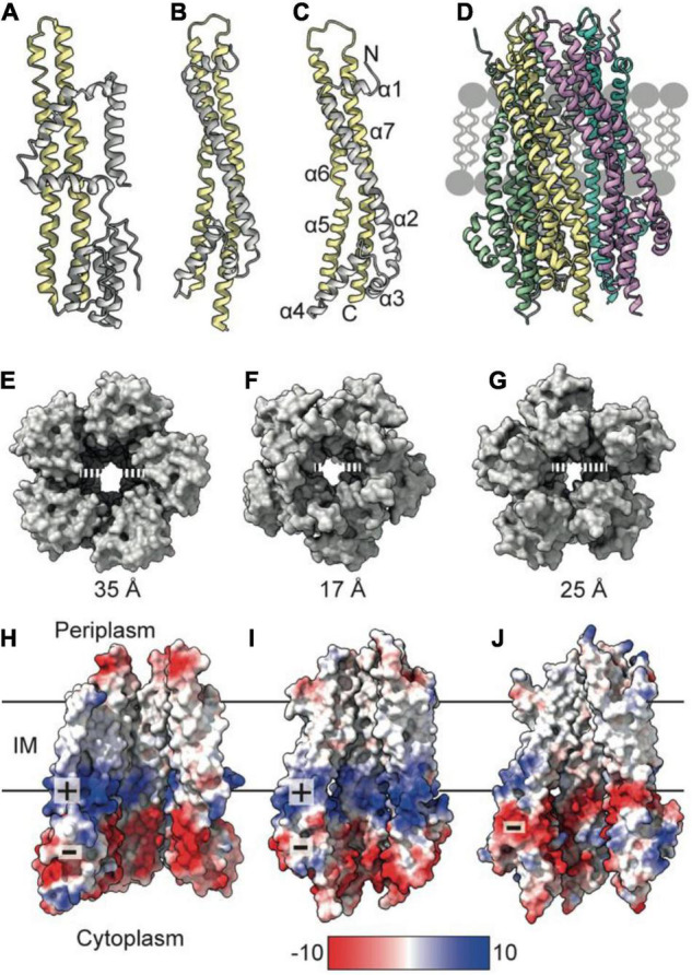
FIGURE 1.

TolQ forms a pentameric pore. (A) MotA monomer transmembrane helices pack together to form a pentameric pore (yellow) and accessory helices are located on the exterior of the pore (gray). (B) ExbB monomer colored as in panel (A). (C) The TolQ monomer (E. coli AlphaFold model) colored as in panel (A), adopts a structure like that observed for ExbB. (D) Pentameric TolQ complex was generated following symmetry expansion of the AlphaFold monomer and energy minimization using RosettaRelax. Each monomer is colored separately. (E–G) The MotA, ExbB, and TolQ pentamers viewed from the cytoplasm to show the outward movement of helices that generate an expanded pore. By contrast, the structures can be overlaid almost perfectly at the periplasmic side. (H–J) A cut through the electrostatic surface of the MotA, ExbB, and TolQ pentamers, respectively, showing that the cytoplasmic side of the pore has a region of negative charge that is conserved across all three systems. A band of positive charge that spans the center of the pore is only observed in MotA and ExbB.
