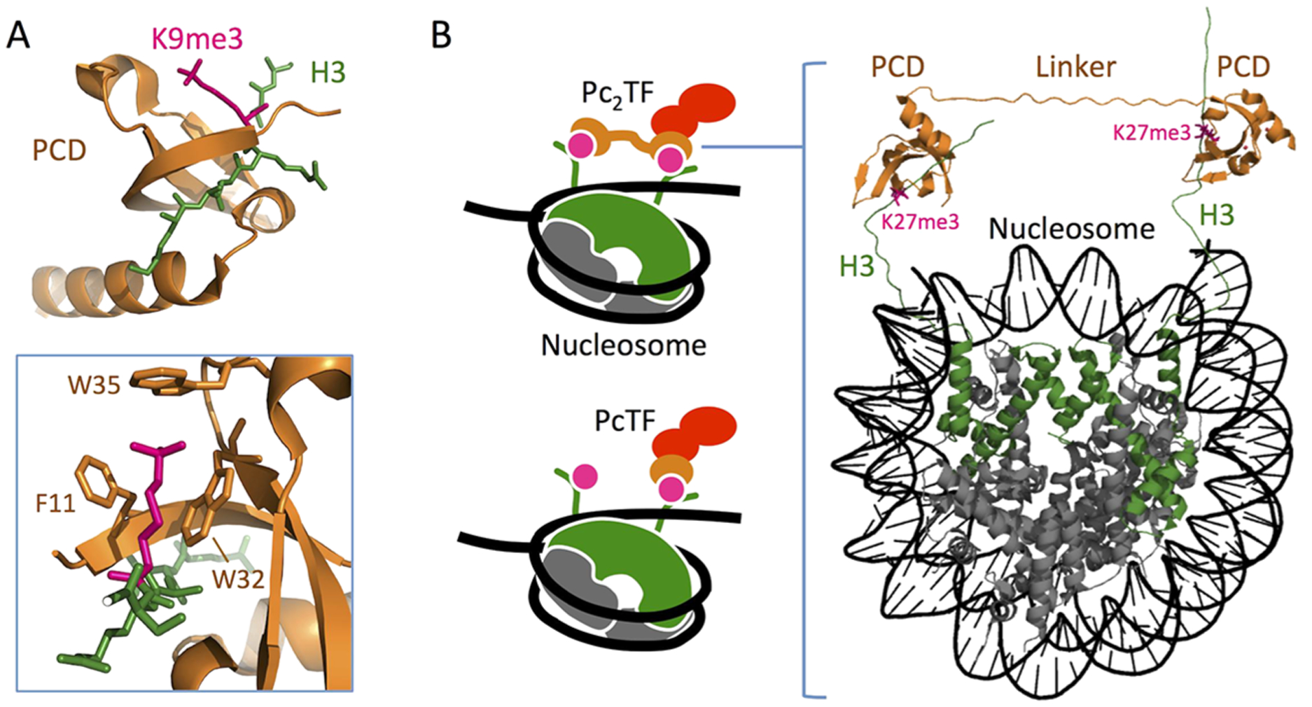
Figure 1.

Three-dimensional model layout to show the plausibility of Pc2TF binding to adjacent H3K27me3 marks. (A) PCD (CBX8) in complex with trimethyl lysine (PDB 3I91).37 Three residues form a hydrophobic cage and surround the Kme3 moiety (inset). (B) H3K27me3 recognition by synthetic fusion proteins that carry a single or tandem PCD domains (PcTF and Pc2TF, respectively). The 3D rendering was composed in the PyMOL Molecular Graphics System, version 1.3, Schrödinger, LLC (https://www.pymol.org), using data for CBX8/H3K9me3 (PDB 4X3K)51 and a whole nucleosome assembly (PDB 5AV8)51,52 from the Protein Data Bank.
