Figures 1–8.
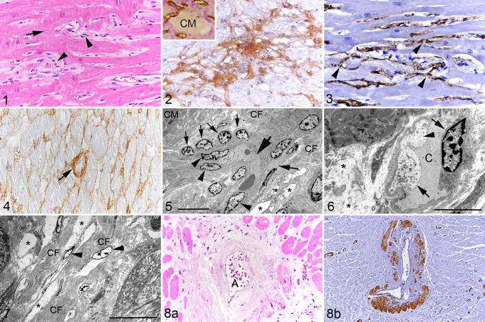
Hypertrophic cardiomyopathy, left ventricular free wall, cat. Figure 1. Case 8. There is widening of the interstitium with increased numbers of interstitial cells (arrowheads) and cardiomyocyte disarray (arrow). Hematoxylin eosin (HE). Figure 2. Case 13. The widened interstitium contains abundant collagen I. Immunohistochemistry (IHC). Inset: Collagen deposition (pink) is present in the interstitium between blood vessels (brown). IHC for CD31 (microvasculature) with van Gieson counterstain. CM: cardiomyocyte. Figure 3. Case 6. Myocardial capillaries are irregularly arranged and appear to branch frequently (arrowheads). IHC for CD31. Figure 4. Case 12. The interstitium contains fine strands of collagen IV. Occasional small capillaries exhibit thickening of the basement membrane due to abundant collagen IV deposition (arrow). IHC. Figure 5. Case 13. Interstitium with a capillary forming a tight curve (large arrow). There is also evidence of branching (medium sized arrow on the right). The nuclei of the lining endothelial cells are relatively plump (arrowheads). Aligned with the capillary is a row of plump pericytes (small arrows). The interstitium is expanded by edema fluid (represented by electron-lucent, amorphous material filling well-demarcated intercellular spaces; asterisks) and irregularly arranged collagen fiber bundles (CF). CM: cardiomyocyte. Transmission electron microscopy (TEM); bar = 10 µm. Figure 6. Case 15. Interstitial capillary (C) with narrowing of the lumen, lined by swollen endothelial cells (arrows) with irregular luminal surface and fluid accumulation in the cytoplasm (arrowhead). The interstitium is distended (edema; asterisks). TEM; bar = 5 µm. Figure 7. Case 12. The interstitium is severely expanded due to abundant collagen fiber bundles (CF) and edema fluid containing amorphous electron-lucent material (asterisks). There are some embedded fibroblasts (arrowheads). TEM; bar = 20 µm. Figure 8. Case 5. Arteriosclerosis of a myocardial artery. (a) Affected artery (A) with diffuse thickening of the tunica media by eosinophilic amorphous material (collagen). HE. (b) Deposition of the proteinaceous material is associated with focal loss of smooth muscle cells in the tunica media and narrowing of the arterial lumen. IHC for α-smooth muscle actin.

