Figures 9–16.
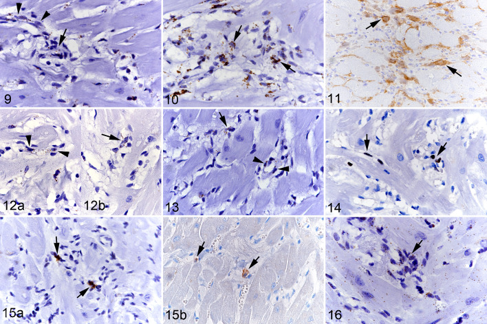
Hypertrophic cardiomyopathy, left ventricular free wall, cat. Figure 9. Case 11. CD34 mRNA expression is seen in the cytoplasm of capillary endothelial cells (arrowheads) and a large proportion of interstitial cells, some of which are arranged in rudimentary ring-shaped structures reminiscent of primitive vessels (arrow). RNA-in situ hybridization (RNA-ISH). Figure 10. Case 8. A large proportion of interstitial cells express Col1A1 mRNA the cytoplasm (arrows). RNA-ISH. Figure 11. Case 13. A moderate number of interstitial cells are positive for procollagen I protein (arrows). Immunohistochemistry (IHC). Figure 12. Case 11. VEGFR2 mRNA expression is seen in the cytoplasm of capillary endothelial cells (arrowheads; [a]), and in several interstitial cells some of which are arranged in rudimentary ring-shaped structures reminiscent of primitive vessels (arrow; [b]). RNA-ISH. Figure 13. Case 11. PDGFRB mRNA expression is seen in the cytoplasm of cells surrounding capillary endothelial cells (pericytes; arrowheads), and in several interstitial cells some of which are arranged in rudimentary ring-shaped structures reminiscent of primitive vessels (arrow). RNA-ISH. Figure 14. Case 8. A few round and spindle-shaped cells in the interstitium express the proliferation marker Ki67 in the nucleus (arrows). IHC. Figure 15. Case 8. (a) Scattered individual cells in the interstitium express Kit mRNA in the cytoplasm and nucleus (arrows). RNA-ISH. (b) Rare interstitial cells show Kit protein expression (arrows). IHC. Figure 16. Case 8. Almost all interstitial cells show a positive signal for MEF2C mRNA in the cytoplasm (arrow). This is also seen in cardiomyocytes. RNA-ISH.

