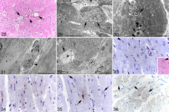
Figures 28–36.
Hypertrophic cardiomyopathy, left ventricular free wall, cat. Figure 28. Case 13. Individual degenerating cardiomyocytes showing sarcoplasmic vacuolation (arrows), hypereosinophilia, and vacuolated and hypereosinophilic sarcoplasm (arrowhead). Hematoxylin eosin (HE). Figure 29. Case 12. Degenerating cardiomyocyte. Mitochondria exhibit diffuse swelling (arrows). C: interstitial capillary. Asterisk: interstitial edema. Transmission electron microscopy (TEM); bar = 20 µm. Figure 30. Case 16. Cardiomyocytes with cytoplasmic lamellar bodies (arrows). TEM; bar = 5 µm. Figure 31. Case 13. Degenerate cardiomyocyte (asterisk) with loss of organelles. The cytoplasm contains fluid and electron-dense debris. C: interstitial capillary with plump endothelial cells and irregular basement membrane. Arrowhead: pericyte. TEM; bar = 20 µm. Figure 32. Case 13. Cardiomyocyte with enlarged nucleus with finely dispersed chromatin (arrow). C: interstitial capillary. TEM; bar = 10 µm. Figure 33. Case 5. Cardiomyocytes exhibit moderate MEF2C mRNA signals. Individual cardiomyocytes exhibit an enlarged nucleus (arrowhead). RNA-ISH. Inset: consecutive section, showing a cardiomyocyte with an enlarged nucleus (arrowhead). Figure 34. Case 5. Cardiomyocytes exhibit strong CD29 mRNA signals. RNA-ISH. Figure 35. Case 5. Cardiomyocytes exhibit a variable Kit mRNA signal (arrows). RNA-ISH. Figure 36. Case 5. Several cardiomyocytes show Kit protein expression (arrows). Immunohistochemistry.

