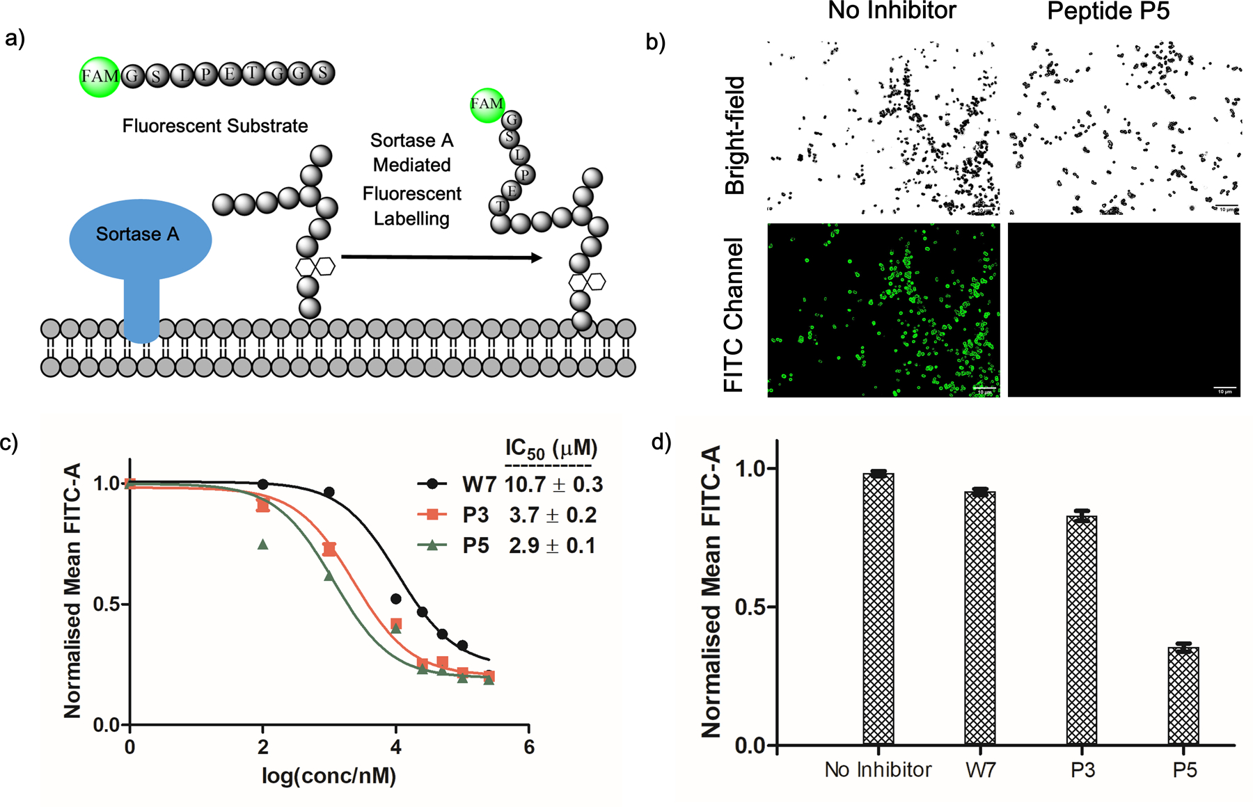
Figure 5.

SrtA inhibition on live S. aureus cells. (a) Schematic representation of SrtA-mediated fluorescence labeling of S. aureus cells. (b) Fluorescence microscopy images showing SrtA inhibition by peptide P5 on live cells, leading to diminished cell staining. P5 concentration, 250 µM; Scale bar, 10 µm. (c) Concentration profiles of W7, P3 and P5 for SrtA inhibition showing the enhanced potency resulting from the reversible covalent warheads. (d) Comparative studies showing that, after washing out the unbound inhibitors, the covalently bound P5 sustained SrtA inhibition over the time course of 6 hours. In contrast, marginal inhibition was observed for W7 and P3 under the same experimental conditions.
