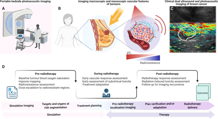
Figure 2.
The potential role of photoacoustic imaging in the clinical radiotherapy framework. (A) Portable bedside PAI can be employed in-room before and/or after RT fractions due to its accessibility, portability and fast acquisitions. (B) PAI could map vascular features of tumors across scales, including blood oxygen saturation. (C) Dual ultrasound and PAI systems provide combined anatomical and molecular imaging features. (D) PAI could be introduced in the clinical RT framework pre-treatment, for diagnostics and pre-operative patient stratification, or for predictive imaging in parallel with CT simulation for radiation dose modulation. During radiotherapy, PAI could be used for monitoring response in the treatment room. After radiotherapy, PAI could further monitor tumor response based on blood oxygen saturation evaluations, which have been associated with local tumor control. PAI could also provide information for response assessment and insights into radiation-induced toxicity at early timepoints. Panel (C) provided in kind by Dr. Oshaani Abeyakoon. Created with BioRender.

