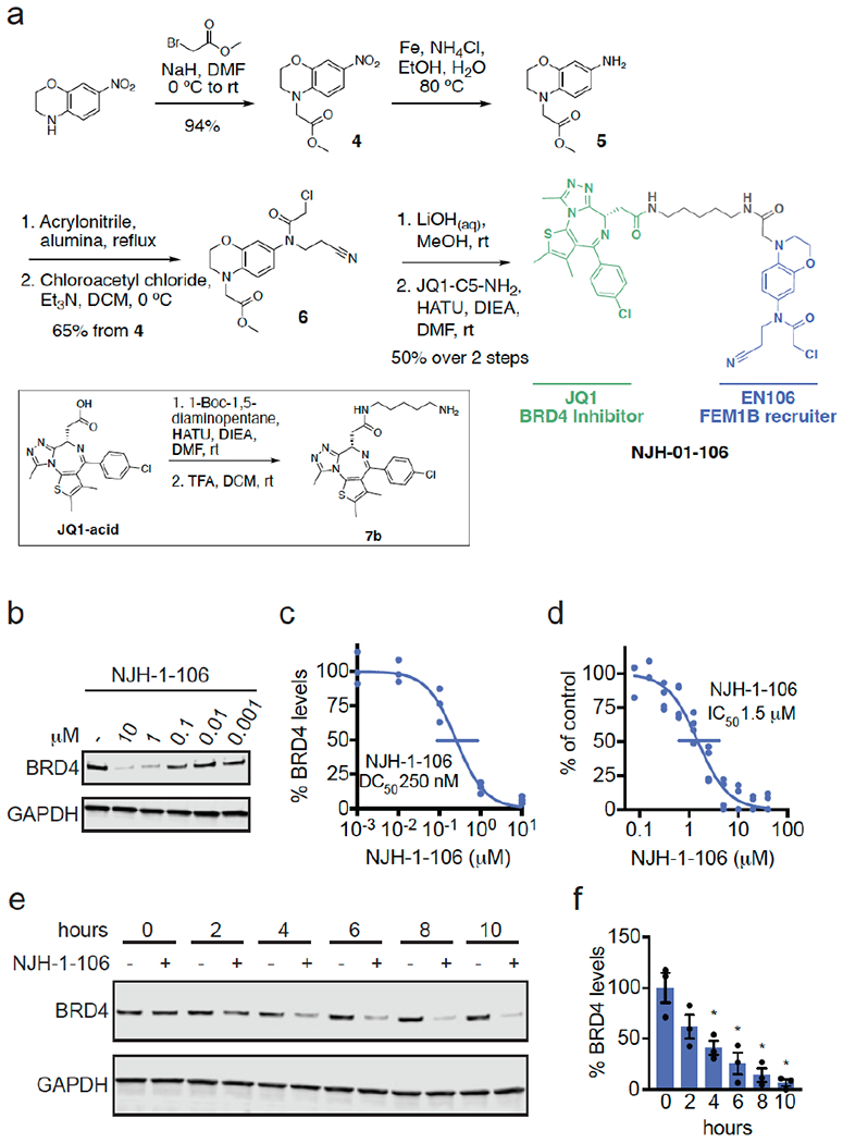
Figure 3. FEM1B-based BRD4 degrader.

(a) Synthesis of FEM1B-based BRD4 degrader NJH-1-106 linking EN106 to JQ1. (b) Degradation of BRD4 by NJH-1-106. NJH-1-106 was treated in HEK293T cells for 8 h and BRD4 and loading control GAPDH levels were detected by Western blotting. (c) Quantification of BRD4 degradation from experiment in (b) and 50 % degradation concentration value (DC50). Individual biological replicate values shown in (b) from n=2-4/group. Gel shown in (b) is representative of n=3 / group which are shown in (c). (d) Dose-response of NJH-1-106 inhibition of FEM1B and TAMRA-conjugated FNIP1 interaction assessed by fluorescence polarization expressed as percent fluorescence polarization compared to DMSO vehicle-treated control. (e) Time course of BRD4 degradation with NJH-1-106 treatment. HEK293T cells were treated with DMSO vehicle or NJH-1-106 (10 μM) for 0, 2, 4, 6, 8, and 10 h and BRD4 and loading control GAPDH levels were detected by Western blotting. Gel shown is representative of n=3 / group. (f) Quantification of BRD4 degradation shown in bar graph as average ± sem with individual biological replicate points shown from experiment designed in (e). Data shown in (c, d, f) are averages with sem in (f) and individual biological replicate values from n=2-4/group
