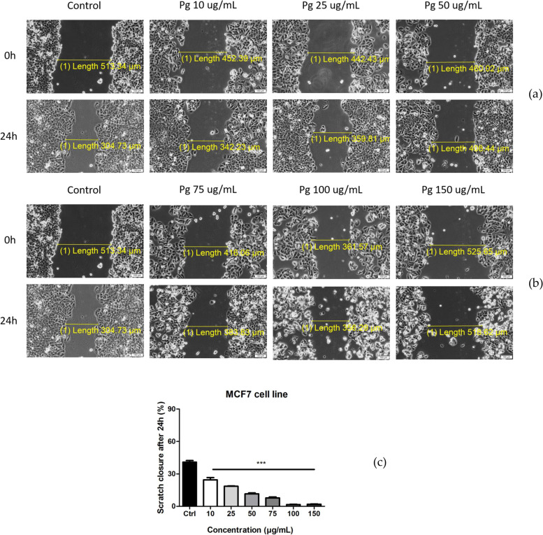
Fig. 7.
a Pg extract (10, 25, and 50 μg/mL) activity on MCF-7 human breast adenocarcinoma cells migration and proliferation potential. Progression of cell migration was monitored by imaging the scratch line initially and at 24 h post-stimulation. Images were taken by light microscopy at 10× magnification; b Pg extract (75, 100, and 150 μg/mL) activity on MCF-7 human breast adenocarcinoma cells migration and proliferation potential. c The anti-migratory potential of Pg extract (10, 25, 50, 75, 100 and 150 μg/mL) on MCF-7 breast adenocarcinoma cells. The bar graphs are expressed as percentage of scratch closure after 24 h compared to the initial surface. Comparison among groups was made using One-way ANOVA test and Dunnett’s multiple comparison post-test. (*** p < 0.001 vs. Control)

