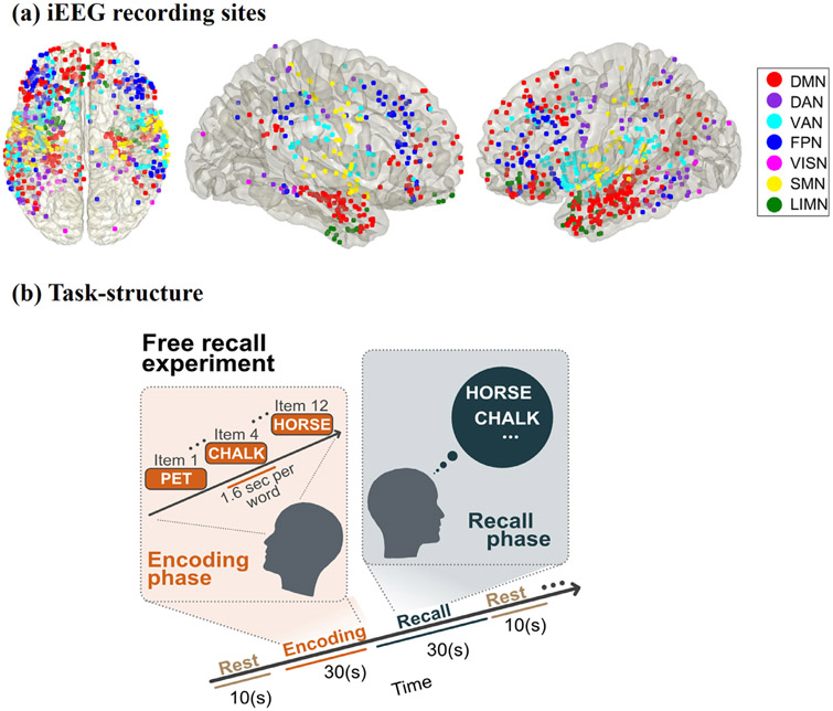
Fig. 1.
(a) iEEG recording sites for the 7 fMRI-derived brain networks investigated in this study. The Yeo cortical atlas was used to map the default mode (DMN), dorsal attention (DAN), ventral attention (VAN), frontoparietal (FPN), visual (VISN), somatomotor (SMN), and limbic (LIMN) networks. In addition to cortical areas, the DMN also included hippocampal regions determined using the Brainnetome atlas (Figs. S1, S2). (b) Cognitive task structure. Participants performed multiple trials of a “free recall” experiment, where they were first presented with a list of words and later asked to recall as many as possible from the original list (see Methods for details).

