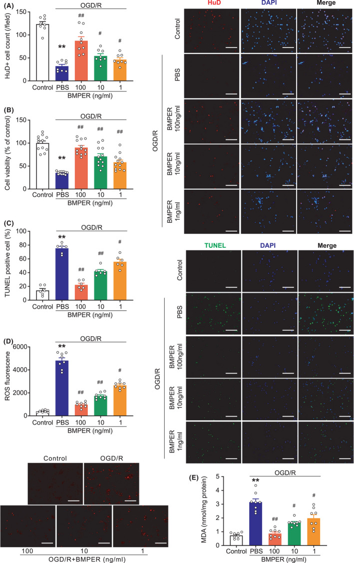
FIGURE 5.

BMPER protects neuronal survival in the OGD/R model. (A) Immunofluorescence staining of live neurons using antibody against HuD, a neuronal marker. DAPI was used to stain nuclei. n = 8. (B) Cell viability of primary neurons subjected to OGD/R stress. n = 12. (C) Immunofluorescence TUNEL showing apoptosis in primary neurons subjected with OGD/R stress. n = 6. (D) ROS production determined by DHE immunofluorescence staining. n = 8. (E) Lipid oxidative stress was evaluated by MDA content. n = 8. Data are expressed as mean ± SEM. **p < 0.01 versus control; # p < 0.05, ## p < 0.01 versus PBS by ANOVA followed by LSD‐t post hoc test. Scale bar = 100 μm. BMP, bone morphogenetic protein; BMPER, BMP‐binding endothelial regulator; DAPI, 4′,6‐diamidino‐2‐phenylindole; DHE, dihydroethidium; MDA, malondialdehyde; OGD/R, oxygen‐glucose deprivation/reperfusion; PBS, phosphate buffered saline; ROS, reactive oxygen species
