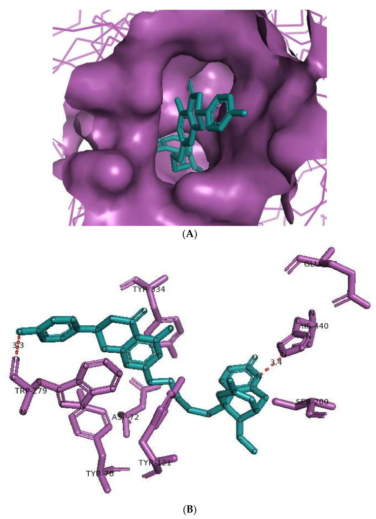
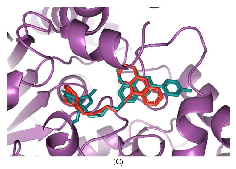
Figure 2.
Docking models of huperzine-4C-naringenin (colored cyan) binding to the active site of AChE enzyme (colored purple). (A) Binding pose of huperzine-4C-naringenin looking down the catalytic gorge of AChE. (B) Ligand huperzine-4C-naringenin docked with the binding site residues of AChE. Hydrogen bonding is represented by red lines and the distance measured in Å. (C) Huperzine-4C-naringenin superimposed with a bis-tacrine dimer (colored red) pre-bound into the crystal structure of the AChE enzyme target (PDB ID: 2CMF).


