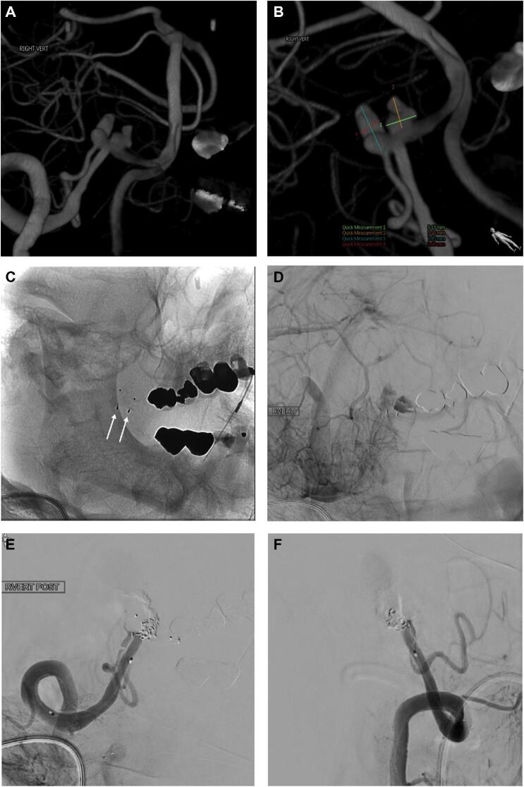
Figure 3.
Patient who had a Hunt and Hess grade III SAH and had intraoperative aneurysm rupture. A and B show a 3D model of a right vertebral artery (VA) injection and 2 dysplastic aneurysms at the origin of the PICA. B, Measurements of both aneurysms are shown in the bottom half of the picture and Table (patient #7). C, Nonsubtracted view showing 2 WEB devices with one in each aneurysm (white arrows). D, Subtracted view in the venous phase after right VA injection showing stasis in both aneurysms. Right VA injection in AP E and lateral F views after deconstruction with Onyx and coils. Note patency of the right PICA on both views.

