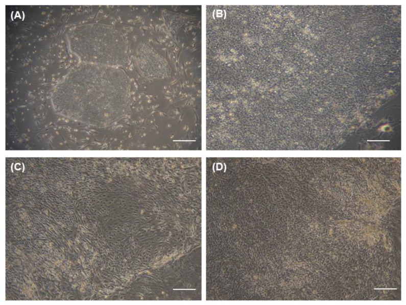
Figure 3.
Morphologic changes of cultured human iPS cells. Bright-field microscopy images of undifferentiated human iPS cell cultures at (A) low-magnification (10×); scale bar = 500 μm) and (B) high-magnification (100×). Bright-field microscopy images of human iPS cells differentiated in (C) limbal-specific (PI) medium only (100×) and (D) PI medium with the optimal BMP4 treatment (100×). Scale bar = 50 μm (B–D).

