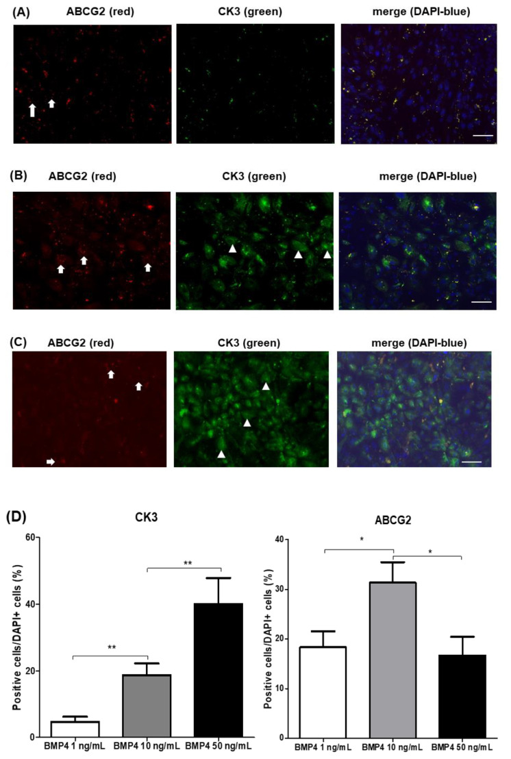
Figure 5.
Immunohistochemical analysis of human iPS cells, differentiated with indicated concentrations of BMP4. Limbal-specific medium with 10 ng/mL BMP4 induced more limbal progenitor cells (ABCG2+, arrow) and less corneal epithelial cells (CK3+, arrowhead) on the differentiation from human iPS cells (B) than 1 ng/mL (A) or 50 ng/mL (C) BMP4. Images represent 3 or 4 independent experiments. Scale bar = 10 μm. (D) Graphs represent ≥ 3 independent experiments, and data are presented as means ± standard deviation (* p < 0.05; ** p < 0.01).

