
Figure 6.
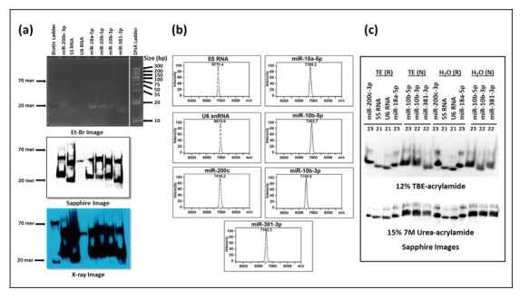
Biotinylation of oligonucleotide primers results in the formation of higher molecular weight species. (a) Analysis of the biotinylated primers on 12% TBE non-denaturing acrylamide gels stained with ethidium bromide (top panel) followed by their transfer onto nylon membranes, incubation with horse-radish peroxide (HRP)-linked streptavidin, and test of the HRP activity using luminol substrate. The chemiluminescent signal was detected either by the Sapphire image analyzer (middle panel) or X-ray development (bottom panel). (b) MALDI-TOF mass spectrometric analysis of the synthesized oligonucleotides provided by the manufacturer. (c) Test of biotinylated primers after heating (95 °C for 5 min followed by cooling on ice). The denatured oligos were analyzed on 12% TBE non-denaturing acrylamide gels (top panel) or 7 M urea/15% acrylamide denaturing gels followed by processing and signal detection as described in panel (a). Detection of the signal was by the Sapphire image analyzer. R = RNA loading buffer with formamide (denaturing); N = Native, non-denaturing loading buffer.