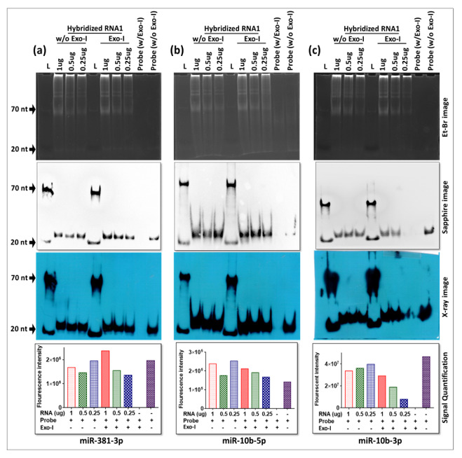
Figure 9.

Successful detection of three miRNAs using liquid hybridization after probe purification. The assay was used to detect expression of: (a) miR-381, (b) miR-10b-5p, and (c) miR-10b-3p. Serial dilutions of RNA (1.00, 0.50 and 0.25 μg each) were subjected to hybridization with the same amount of gel purified biotinylated oligos (2.5 pmol/reaction). The hybridized mixtures either without (w/o) or with (w) Exo-I treatment were loaded on 15% non-denatured acrylamide-TBE gel followed by transfer on a nylon membrane(s) using a semi-dry blotter. Gels were stained with ethidium bromide for RNA visualization under UVP. After transfer, each nylon membrane was subjected to UV crosslinking and incubation with HRP-conjugated streptavidin before detection by the Sapphire imaging system or X-ray. After transfer, membranes were probed with pre-biotinylated miR-381-3p, miR-10b-5p and miR-10-3p probes, respectively, and expression of these miRNAs was detected using X-ray. Quantitative analysis of expression levels of miRNAs was performed using GelQuant.NET software. Relative % intensities were measured for each mRNA and plotted against the corresponding amount of RNA.
