
Figure 12.
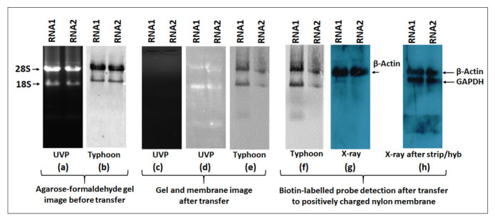
Inability of Typhoon imaging to detect the ECL signal compared to X-ray imaging. RNA1 and RNA2 were subjected to electrophoresis on 1.2% denatured agarose-formaldehyde gel. (a) The gel image was taken with a UVP gel doc system after ethidium bromide (EtBr) staining, but before transfer to positively charged nylon membranes. (b) The gel image was taken with the Typhoon imaging system before transfer to positively charged nylon membranes. (c) The gel image was taken after transfer to a nylon membrane by UVP gel doc. (d) The nylon membrane image was taken by UVP after transfer. (e) The nylon membrane image was taken by Typhoon after transfer using the EtBr filter. (f) Detection of a biotin-labeled probe by Typhoon using ECL filter. (g) Detection of β-actin biotin-labeled probe by X-ray. (h) Detection of GAPDH biotinylated probe by X-ray after mild stripping of the blot shown in panel (g) after hybridization with β-actin-biotinylated probe.