Figure 2.
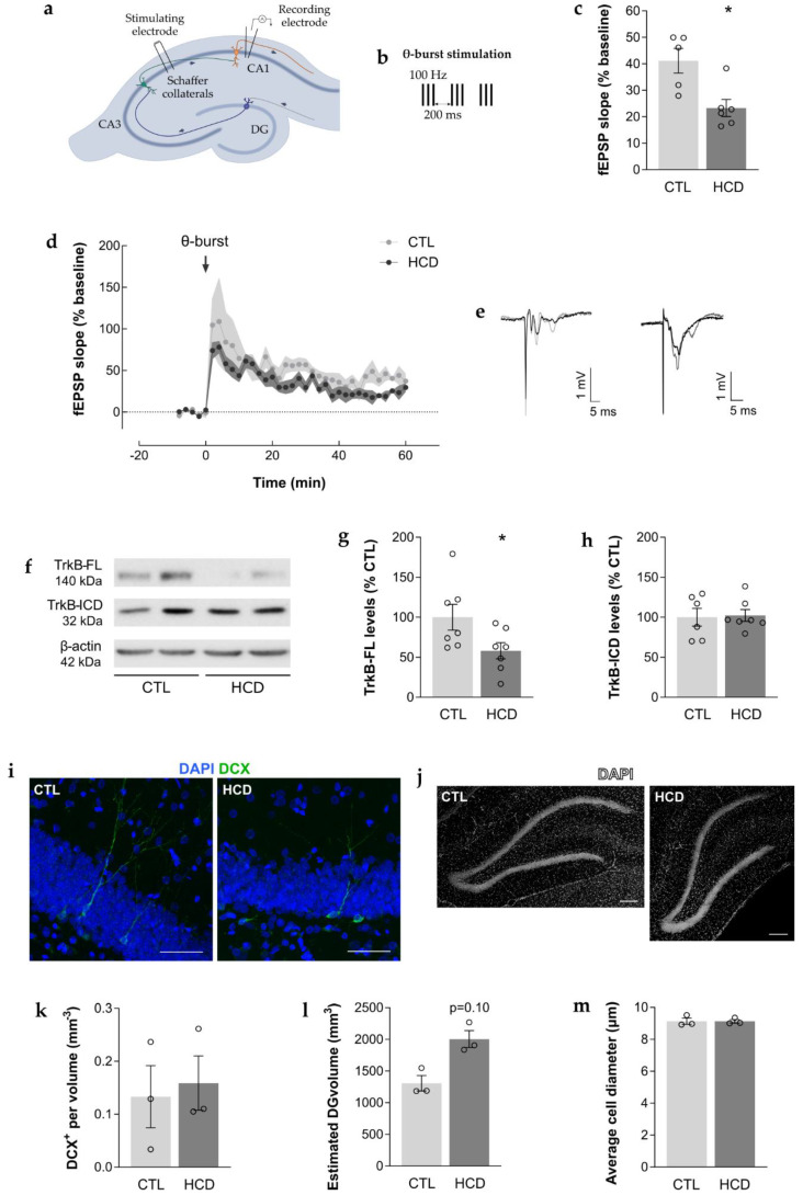
A chronic high caloric diet (HCD) promoted hippocampal synaptic plasticity impairment and reduced the levels of hippocampal TrkB-FL without impacting neurogenesis. (a,b) Representative scheme of electrophysiological recordings in acute hippocampal slices. (a) Schematic representation of an acute hippocampal slice with the electrophysiological recording configuration used to obtain field excitatory postsynaptic potentials (fEPSPs) from the CA1 area under the stimulation of Schaffer collateral/commissural fibers in the stratum radiatum of the CA1 area. (b) Representation of the applied long-term potentiation (LTP) protocol. After a stable baseline (10 min), LTP was induced through a weak θ-burst protocol (3 trains of 100 Hz, 3 stimuli, separated by 200 ms). (c–e) Hippocampal synaptic plasticity of 24-month-old HCD rats was impaired, as assessed by electrophysiological extracellular recordings. (c) Significant decrease in LTP magnitude, quantified as the fEPSP average slope (% baseline) obtained between 50 and 60 min after LTP induction (θ-burst). * p < 0.05, unpaired Student’s t-test. Data are expressed as mean % ± SEM. (d) The averaged time-course changes in fEPSP slope (% baseline) induced by a θ-burst stimulation. Data are expressed as mean % ± SEM. (e) Tracings from representative experiments. For each condition, fEPSP tracings recorded at baseline (baseline, grey line) and after θ-burst-induced LTP (LTP, black line) from the same slice are shown overlaid. (f–h) Levels of TrkB full length (TrkB-FL) were reduced in the hippocampus of 24 month old HCD rats, as assessed by western blot (WB). (f) Representative WBs depict immunoreactive bands for TrkB-FL (~140 kDa), TrkB-ICD (~32 kDa) and β-actin (loading control, ~42 kDa). (g,h) Protein levels were quantified and normalized (100%) for the corresponding controls (% CTL). (g) Significant decrease in the levels of TrkB-FL, * p < 0.05, unpaired Student’s t-test, which did not result in (h) changes in the levels of TrkB intracellular domain fragment (TrkB-ICD), p > 0.05, unpaired Student’s t-test. (i–m) The density of immature neurons was not altered in the dentate gyrus (DG) of the hippocampus of 24-month-old HCD rats, as assessed by IHC. (i) Representative coronal sections immunostained with 4′,6-diamidino-2-phenylindole (DAPI) (blue) and doublecortin (DCX) (green). Scale bar = 50 μm. (j) Representative coronal sections immunostained with DAPI (white). Scale bar = 200 μm. (k) No significant difference in the density of DCX+ cells. * p > 0.05, Mann–Whitney test. (l) No significant differences in the estimated volume of the DG or (m) in the average diameter of DAPI+ cells. p > 0.05, unpaired Student’s t-test. (c,d) Data are expressed as mean % ± SEM. (g,h,k–m) Data are expressed as mean ± SEM.

