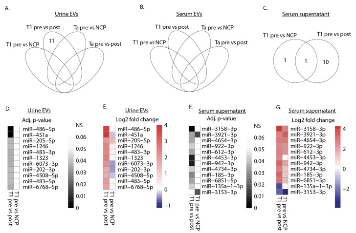
Figure 2.
Differential expression of miRNAs in urine EVs and serum supernatant. (A–C) Venn diagram depicting the number of DEmiRNAs in presurgery (pre) samples compared to postsurgery (post) samples and NCPs in urine EVs, serum EVs, and serum supernatant. (D,E) Heat map displaying adjusted p-values and log2 fold change values for the eleven DEmiRNAs detected in (A) urine EVs. miRNA expression levels were compared between T1 presurgery samples and postsurgery samples or NCPs. (F,G) Heat map displaying adjusted p-values and log2 fold change values for the twelve DEmiRNAs detected in (C) serum supernatant. miRNA expression levels were compared between T1 presurgery samples and postsurgery samples or NCPs. An adjusted p-value threshold of 0.05 was set to determine DEmiRNAs for all analyses and non-significance (NS) is denoted in the scale.

