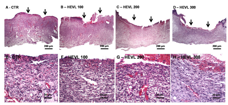
Figure 3.
Photomicrographs of histological sections stained in HE representative of the healing wound area in Wistar rats of the experimental in day 7. (A–D) Panoramic view of CTR and treated with HEVL at doses of 100, 200 and 300 mg/Kg, respectively. Note the non-epithelized wound surfaces (black arrows) (40×). (E) CTR shows immature vascular and edematous granulation tissue, whereas (F) HEVL 100 and (G) 200 presents greater content of proliferative spindle-shape cells (400×). (H) In HEVL 300, granulation tissue was less vascular and more cellular (400×).

