Figure 4.
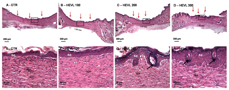
Photomicrographs of HE-stained histological sections representative of the healing wound area in Wistar rats from different groups after 14 days of experiment. (A–D) Histological panoramic view highlighting the area of scar repair in groups CTR and treated with HEVL at doses of 100, 200 and 300 mg/Kg, respectively. Note the presence of full re-epithelialization of the wound surface (red arrows), as well as the diameter of the CTR healing area considerably larger than in the HEVL-treated groups (thin double-headed arrows) (40×). Higher magnifications show thin keratinized squamous epithelium with no morphological signs of cutaneous appendages differentiation (E) CTR and (F) HEVL 100 (HE, 400×). (G) Group HEVL 200 and (H) HEVL 300 exhibiting bulbous buds, most of them with central keratinization, compatible with hair follicles (400×). Black arrows in G and H point at the neoformation of cutaneous appendages (hair follicles). The fibrous connective tissue (TCF) shows persistence of chronic inflammatory infiltrate in CTR whereas inflammation was inconspicuous in the other groups.

