Figure 5.
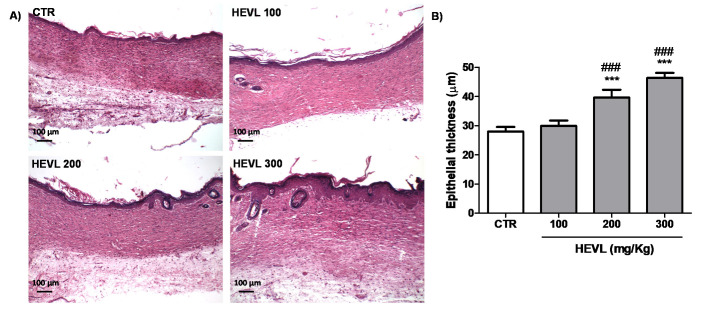
(A) Photomicrographs of histological sections stained in HE representative of the scarring tissue in the healing wound area of the experimental in day 14. The scar area is lined by keratinized squamous epithelium with presence of rudimentary hair follicles in HEVL 200 and well-developed hair follicles in HEVL 300 (100×). (B) Assessment of the epithelial thickness in the experimental groups on day 14. Data are expressed as median, interquartile range and maximum and minimum values. Significant differences in relation to the CTR (control) group are expressed as *** p <0.001; significant differences in comparison with HEVL 100 are expressed as ### p < 0.001 (Kruskal-Wallis test and Dunn’s multiple comparison test).

