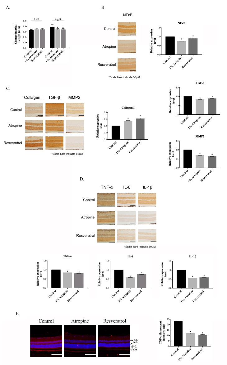
Figure 1.
Effect of resveratrol on myopia progression. (A) The axial length was determined as the difference in diopter measurements taken after and before MFD. * p < 0.05 indicates significant differences from the control. Quantification of immunohistochemical staining for NFκB (B), collagen I, TGF-β, and MMP2 (C), and TNF-α, IL-6, and IL-1β (D). (E) Immunofluorescence staining with TNF-α and quantification analysis of the RPE layer by Image J software. Results are shown as mean ± SD; The scale bar indicates 50 μm, 20× magnification for IHC stain images, 100 μm, 20× magnification for IF stain images * p < 0.05, n = 3 for each.

