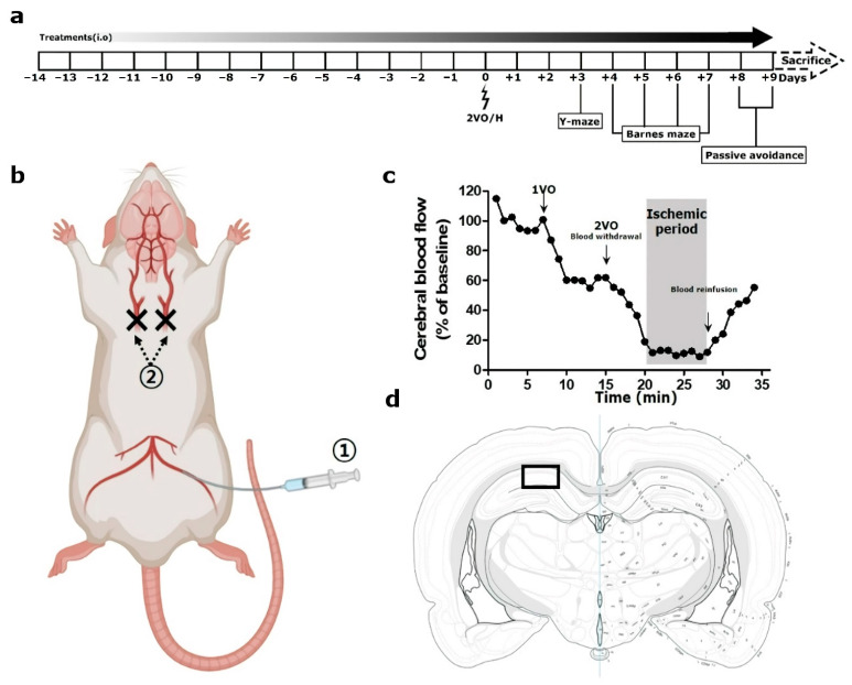
Figure 2.
Graphical illustration of the in vivo experiment setup. (a) Schematic overview of the in vivo experiment, (b) surgical procedure, (c) a representative Doppler flowmetry traced for the entire operation time, and (d) the area of interest for histologic analyses in this study are presented. In (b), circled “1” and “2” represent the steps of femoral artery catheterization and ligatures of both common carotid arteries in the 2VO/H operation, respectively. In (d), a rectangular box indicates a 300 µm-width area in hippocampal Cornu ammonis 1, which was used for histologic analyses.

