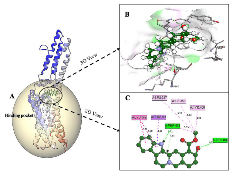
Figure 6.
(A) Homology modeled structure of 5HT1A docked with yohimbine shown in ball and stick representation in green color within the binding site sphere of pink color. (B) Close intra-molecular interactions formed with the best ligand conformation. (C) 2D view of close intra-molecular interactions formed within the binding pocket residues and yohimbine as shown in green color ball and stick presentation, hydrogen bonds are shown in green dashed line, pi-sigma bonds in purple dashed line, and pi-alkyl bonds in pink dashed lines. The diagrams are prepared in Discovery Studio Client V 20.1.

