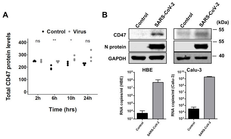
Figure 1.
SARS-CoV-2 infection is associated with increased CD47 levels. (A) TF protein abundance in uninfected (control) and SARS-CoV-2-infected (virus) Caco-2 cells (normalized signal intensity, data derived from [23]). p-values were determined using two-sided Student’s t-test. (B) CD47 and SARS-CoV-2 N protein levels and virus titers (genomic RNA determined by PCR) in SARS-CoV-2 strain FFM7 (MOI 1)-infected air−liquid interface cultures of primary human bronchial epithelial (HBE) cells and SARS-CoV-2 strain FFM7 (MOI 0.1)-infected Calu-3 cells. Uncropped blots are provided in Figure S1. p-values were determined by two-sided Student’s t-test. * p < 0.05, ** p < 0.01.

