Figure 1.
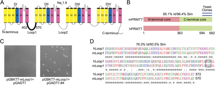
Yeast two-hybrid screen to identify proteins that interacted with NaV1.9. (A) Schematic structure of the NaV1.9 channel. Residues 402 to 570 of mNaV1.9 were used as bait for the yeast two-hybrid (Y2H) screen. (B) Location of the repeat-independent clones of PRMT7 identified by Y2H (blue lines, residues 594-692). The 2 domains (pink/green) and degrees of homology between full-length mouse and human PRMT7 are shown. (C) Residues 594 to 692 of mouse PRMT7 were purified from yeast clones for direct Y2H to confirm the interaction on QDO medium. (D) Amino acid sequence comparison of hLoop1 and mLoop1 using ClustalW (EMBL–European Bioinformatics Institute). Potential PRMT7 methylation motif, RXR, is boxed. PRMT7, protein arginine methyltransferase 7; QDO, quadruple dropout.

