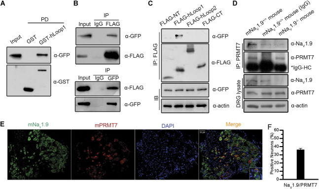
Figure 2.
Interaction and coexpression of NaV1.9 with PRMT7. Interaction between hLoop1 and full-length hPRMT7 was analysed by GST pull-down assays (A) and reciprocal coimmunoprecipitation assays (B). (C) Interaction between the intracellular domains of hNaV1.9 and hPRMT7 was examined by coimmunoprecipitation assays. (D) Interaction of mPRMT7 with mNaV1.9 in mouse DRG tissues was assessed. Scn11a +/+ or Scn11a −/− mouse DRG tissue lysates were immunoprecipitated using anti-mPRMT7 antibodies and control IgG. The precipitates were immunoblotted with the indicated antibodies. (E) Immunohistochemical analysis of mPRMT7/mNaV1.9 expression in mouse DRG sections. The sections were stained for mPRMT7 (red) and mNaV1.9 (green) and DAPI (blue). mPRMT7 showed considerable colocalization with mNaV1.9 in mouse DRG neurons. Scale bars, 50 μm. (F) Percentages of mPRMT7-positive and mNaV1.9-positive neurons are shown. DAPI, 4,6-diamidino-2-phenylindole; DRG, dorsal root ganglion; GST, glutathione S-transferase; IgG, immunoglobulin G; hNaV1.9, human hNaV1.9; PRMT7, protein arginine methyltransferase 7.

