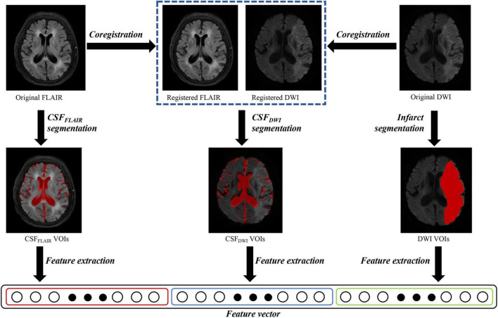
FIGURE 2.
Overview of image processing with diffusion weighted imaging (DWI) and fluid attenuated inversion recovery (FLAIR) images for each patient. Infarct regions were segmented on the DWI images, and the cerebrospinal fluid regions were segmented on the FLAIR images (CSFFLAIR). The FLAIR images were overlaid onto the DWI images, and the cerebrospinal fluid regions on DWI images (CSFDWI) were segmented on the registered DWI images. One thousand three hundred and sixteen features were extracted from DWI volumes of interest (VOIs), CSFFLAIR VOIs and CSFDWI VOIs, respectively.

