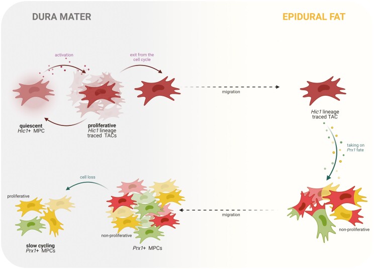
Figure 7.
Hypothetical model proposed for the Hic1 and Prx1 hierarchy. Hic1+ MPCs native to the dura mater proliferate in response to biological cues (Fig. 2D, 2E). These MPCs exit from the cell cycle while in the dura mater (Fig. 2F) and then these Hic1+ lineage traced TACs migrate to the adjacent epidural fat (Fig. 2F). In the epidural fat, these MPCs/TACs acquire a Prx1+ mesodermal fate, as seen by the colocalization of Hic1 and Prx1 expression in cells (Fig. 4; Supplementary Fig. 5). These Prx1+ MPCs migrate back to the dura mater and do not re-enter the cell cycle. The expansion of Prx1+ MPCs in the dura mater over time (Fig. 2A-2C) is a product of the migration of these cells from the epidural fat and not due to cell proliferation. At skeletal maturity, Prx1+ MPCs in the dura mater are lost aside from a few interspaced MPCs, which are now proliferative (Fig. 3) and most likely undergo slow cycling in this tissue to maintain homeostasis and respond to future activation cues/insults (Fig. 6). Abbreviations: Hic1, hypermethylated in cancer 1; MPCs, mesenchymal progenitor cells; Prx1, paired related homeobox-1; TACs, transit-amplifying cells.

