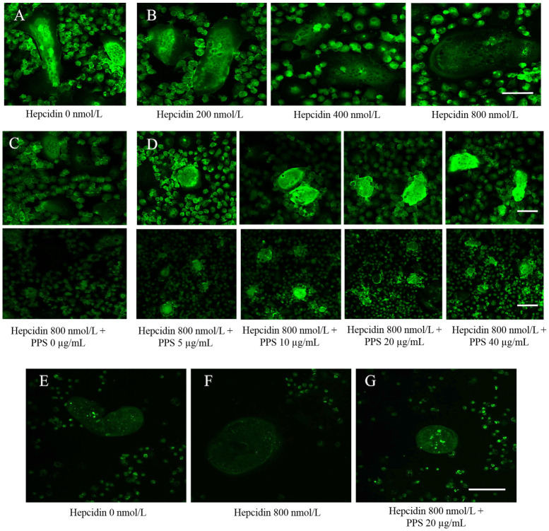
Fig 5. Immunofluorescence images.
(A) Immunofluorescence analysis of ferroportin 1 (FPN1) protein. Canine bone marrow derived OC were treated with rabbit anti-ferroportin 1 after 20 hours treatment of hepcidin (200, 400 and 800 nmol/L) to visualize the expression of FPN1 protein. The figure shows the strong detection of localization of FPN1 at the membrane of hepcidin untreated OC (control). (B) The green fluorescence intensity significantly weakened toward the higher concentration of hepcidin (800 nmol/L). Scale bar- 50 μm. (C) PPS inhibits the hepcidin-induced FPN1 internalization and degradation. OC were treated with rabbit anti-ferroportin 1 after 20 hs treatment of PPS (5, 10, 20 and 40 μg/mL) and hepcidin 1 (800 nmol/L) to visualize the expression of FPN1 protein. The figure shows the strong inhibition of localization of FPN1 at the membrane of hepcidin treated OC (control). (D) Heparin induced FPN1 internalization and degradation was inhibited with higher concentrations of PPS (5, 10, 20 and 40 μg/mL). Higher the fluorescence intensity was visualized toward the 40 μg/mL PPS. Scale bars- 50 and 100 μm. (E) Confocal microscopy analysis of iron concentration in OC. Cell medium was replaced with fresh medium containing 25 ng/mL M-CSF, 50 ng/mL RANKL and 800, nmol/L hepcidin 1 and 20 μg/mL PPS and allowed to incubate for 20 h. Fluorescence of the Phen Green FL is quenched upon binding iron and emission intensity was weakened when the iron concentration is high. E, shows high fluorescence intensity in untreated cells implying low intracellular iron. (F) Fluorescence images showed that intracellular iron concentration was increased (weakened fluorescence intensity) with hepcidin 800 nmol/L compared to the untreated cells. (G) fluorescence images have proven the inhibitory effect of PPS on hepcidin-induced iron accumulation by visualizing greater intensity of fluorescence at 20 μg/mL.

