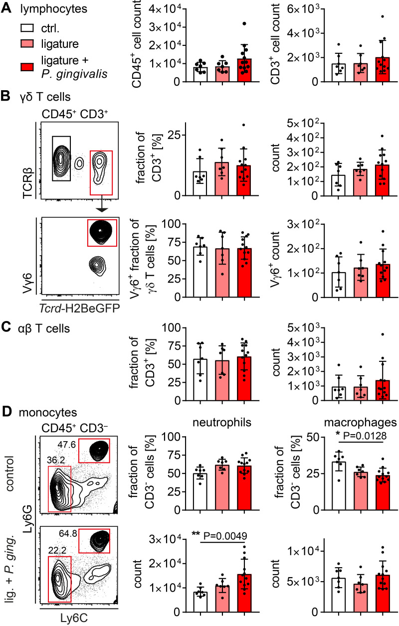
Fig 2. Cellular immune reaction to ligature-induced periodontitis.
(A) Number of CD45+ lymphocytes and CD3+ T cells isolated from gingival tissue of ligature-treated and control mice. (B) Fraction and number of γδ T cells isolated from gingival tissue of ligature-treated and control mice in CD45+ CD3+ or CD45+ CD3+ TCRγδ+ gate, respectively, and representative gating. (C) Fraction and number of αβ T cells isolated from gingival tissue of ligature-treated and control mice in CD45+ CD3+ gate. (D) Fraction and number of neutrophils and macrophages in CD45+ CD3- gate. In all diagrams each point represents a mouse. Data were obtained from three independent experiments and statistically analyzed by Kruskal-Wallis test and Dunn´s multiple comparisons, mean and SD are shown. A p‐value below 0.05 was considered as significant.

