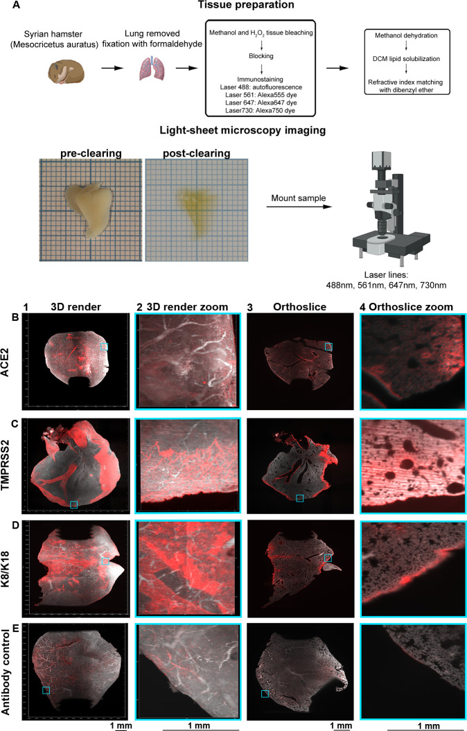
Fig 1. Volumetric analyzes of ACE2 and TMPRSS2 in non-infected Syrian hamster lung lobes delineate expression patterns.
(A) Lung lobes from Syrian hamsters are extracted and fixated in 10% formalin. Tissue preparation is performed with bleaching, followed by blocking, immunostaining, dehydration, lipid solubilization, and refractive index matching. Refractive index matching for increased light penetration (pre-clearing vs. post-clearing on 1 mm paper), after which imaging is performed and data is analyzed. Orthogonal slices are generated with ImageJ and 3D renders with Imaris. Autofluorescence in grey (488 channel) and staining in red (647 channel). (B) ACE2 is distributed over the lung with nonuniform signals in the tertiary bronchi, bronchioles, and alveoli. (C) Significant TMPRSS2 expression in primary, secondary, and tertiary bronchi with minor alveolar presence. (D) Intense K8/K18 staining in the primary and secondary bronchi, with occasional signal in tertiary bronchi, bronchioles, and alveoli. (E) Autofluorescence in the 647 channel and minor staining of the outer regions of the lung for the secondary antibody control, the pattern does not overlap with previous stains.

