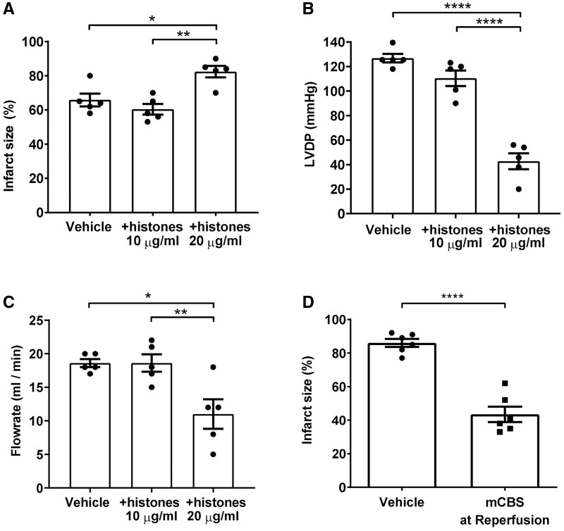
Figure 4.
Extracellular histones contribute to myocardial infarction during reperfusion, and histone neutralization during reperfusion is cardioprotective. (A–C) Exogenous histones or vehicle were added to the perfusate of Langendorff-perfused rat hearts following 45 min ischaemia and during 2 h reperfusion. Infarct size (B) was measured at the end of the experiment. Left ventricular developed pressure (LVDP) (B) and flow rate of the perfusate (C) were measured 5 min into the reperfusion period. N = 5 hearts per group. (D) Infarct size in hearts subject to 45 min of global ischaemia followed by 2 h reperfusion, in the presence of vehicle or 100 μg/mL mCBS. N = 6 hearts per group. Statistical analyses by one-way ANOVA with Tukey post-test (A–C), or by unpaired T-test (D). *P < 0.05, **P < 0.01, ****P < 0.0001.

