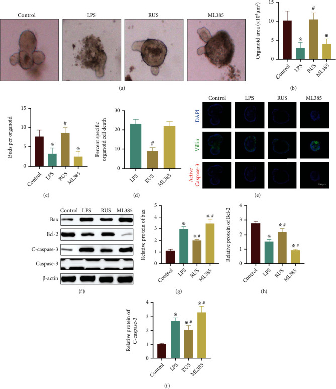
Figure 6.

RUS improved intestinal epithelial barrier function by inhibiting epithelial cell apoptosis. (a) Growth of organoids in the control group, LPS group, RUS group, and ML385 group (with RUS) as observed by a light microscope. (b, c) The size and buds of organoids in each group. (d) MTT assay showing the activity of organoids. (e) Immunofluorescence analysis showing the distribution of Villin (green) and active Caspase-3 (red) in organoids. DAPI (blue) was used to stain the nuclei. (f–i) The protein levels of Bcl-2, Bax, and C-caspase-3 in organoids as determined by WB.✲P < 0.05, compared with the control group. The experiments were repeated 3 times, and the most typical result is shown. Data are presented as the means ± SD (※P < 0.05, compared with the control group; #P < 0.05, compared with the LPS group).
