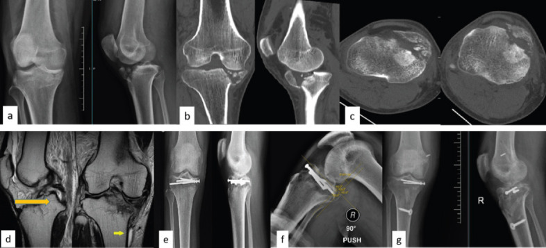
Figure 1.

Case 1: Preoperative X-ray (a) and CT images (b and c) showing large anterior compression fracture of tibia, MRI images showing PCL and MCL tear (d), Postoperative X-ray (e) PCL stress X-ray showing posterior subluxation (f), and Post PCL and MCL reconstruction X-ray (g). PCL: Posterior cruciate ligament, MCL: Medial collateral ligament
