
Figure 2.
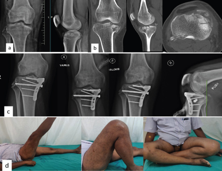
Case 3: Pre-operative X-ray (a) and CT (b) showing large anterior compression fracture of the tibia and fibular head fracture, Post-operative X-rays showing fracture union and stable joint on valgus, varus, and posterior stress ©, Follow-up pictures showing good knee ROM (d).