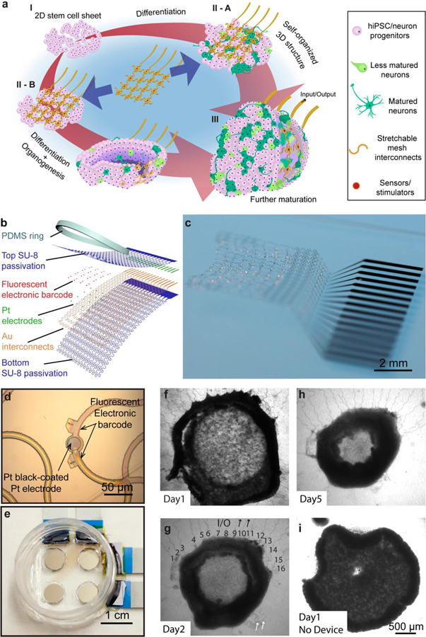
Figure 1. Stretchable mesh nanoelectronics for brain organoid integration.
a, Schematics illustrate the stepwise integration of stretchable mesh nanoelectronics into 3D hiPSC-derived neural tissues through cell self-organization and brain organoids through organogenesis. (I) Human induced pluripotent stem cells (hiPSCs) were seeded with Matrigel. (II) Lamination of stretchable mesh nanoelectronics onto the two-dimensional (2D) cell sheet after neuronal differentiation into neural progenitors (II-A) or lamination of stretchable mesh nanoelectronics onto the hiPSCs before neuronal differentiation (II-B). (III) The 2D-to-3D self-organization folds the 2D cell sheet/nanoelectronics hybrid into a 3D structure. 3D embedded sensors are connected to external recording electronics to keep monitoring the electrophysiology of hiPSC-derived neurons and neural progenitors. b, Exploded view of the stretchable mesh nanoelectronics design consisting of (from top to bottom) an 800-nm-thick top SU-8 encapsulation layer, a 50-nm-thick platinum (Pt) electrode layer electroplated with Pt black, a 40-nm-thick gold (Au) interconnects layer, and an 800-nm-thick bottom SU-8 encapsulation layer. The serpentine layout of interconnects is designed to enable stretchability. A polydimethylsiloxane (PDMS) ring is bonded around the device as a chamber to define the size and initial cell number in the seeded hiPSC sheet. c, Optical photograph of stretchable mesh nanoelectronics released from the substrate and floating in the saline solution. d, Optical bright-field (BF) microscopic image of stretchable mesh nanoelectronics before released from the fabrication substrate shows a single Pt electrode coated with Pt black. e, Optical photograph of a 2×2 devices well, with a single culture chamber for 4 cyborg brain organoids cultured simultaneously. f-h, Optical phase images of hiPSC-derived neurons integrated with stretchable mesh nanoelectronics from Day 1 to Day 5 show that the 2D cell sheet with embedded stretchable mesh nanoelectronics self-folded into a 3D cyborg brain organoid. Black numbers and arrows indicate the input/output (I/O) stretchable connectors for the 16-channel electrode array (g). White arrows highlight the stretchable anchors used to keep the stretchable mesh nanoelectronics unfolded on the substrate, which were released after seeding with cells (g). i, Optical phase images of organoid without nanoelectronics integrated at Day 1 of culture as control showing minimal interruption from the integration with stretchable mesh nanoelectronics to the organogenesis of brain organoids.

