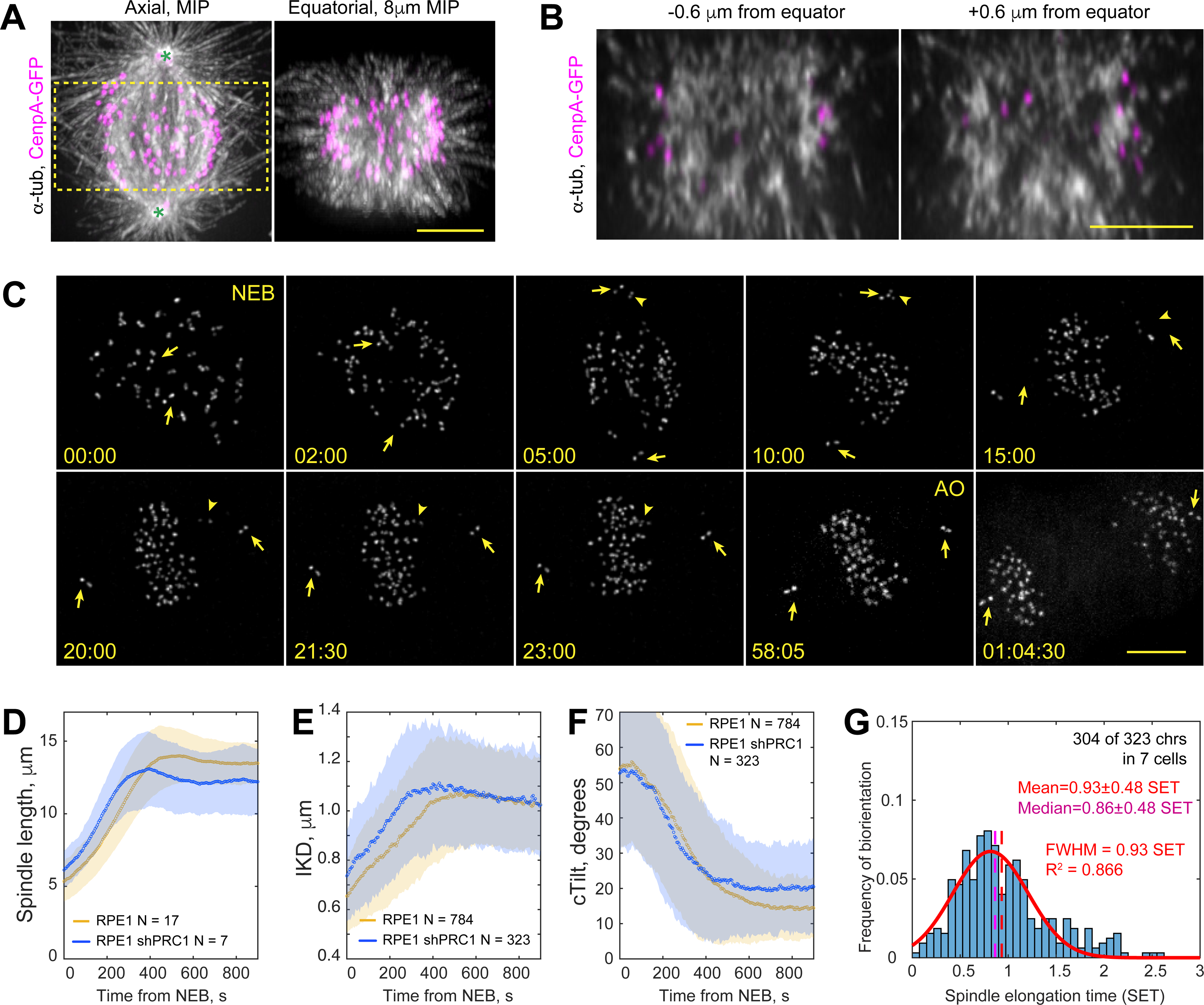
Figure 3. Lack of microtubule bundles delays formation of amphitelic attachments.

(A) Spatial arrangement of MTs (α-Tubulin), KTs (CenpA-GFP), and centrioles (Ctn1-GFP) in a prometaphase shRNA-depleted of PRC1. Axial view is a maximum-intensity projection of the entire spindle. Equatorial view presents a partial volume denoted by the box in Axial view. Asterisks denote centrioles (~12-μm spindle length). (B) Individual equatorial planes from the from the volume shown in (A). (C) Selected timepoints from a recording of PRC1-depleted RPE1 cell with GFP-tagged KTs and centrioles. Frames are maximum-intensity projections of the entire cell. Nuclear envelope breaks down at 00:00 (NEB) and anaphase onsets at 58:05 (AO). Arrows mark centrioles, arrowheads – a monooriented chromosome. (D-F) Dynamics of mean spindle length (D), distance between sister KTs (E, IKD), and the angle between the centromere and spindle axes (F, cTilt). Colored corridors are ±1 STD. (G) Temporal distribution of biorientation events in PRC1-depleted cells, normalized to spindle elongation time. Notice significant deviation from the normal distribution (red line). Scale bars, 5 μm in (A), (B) and (C). See also Figures S2, S3 and Video S3.
