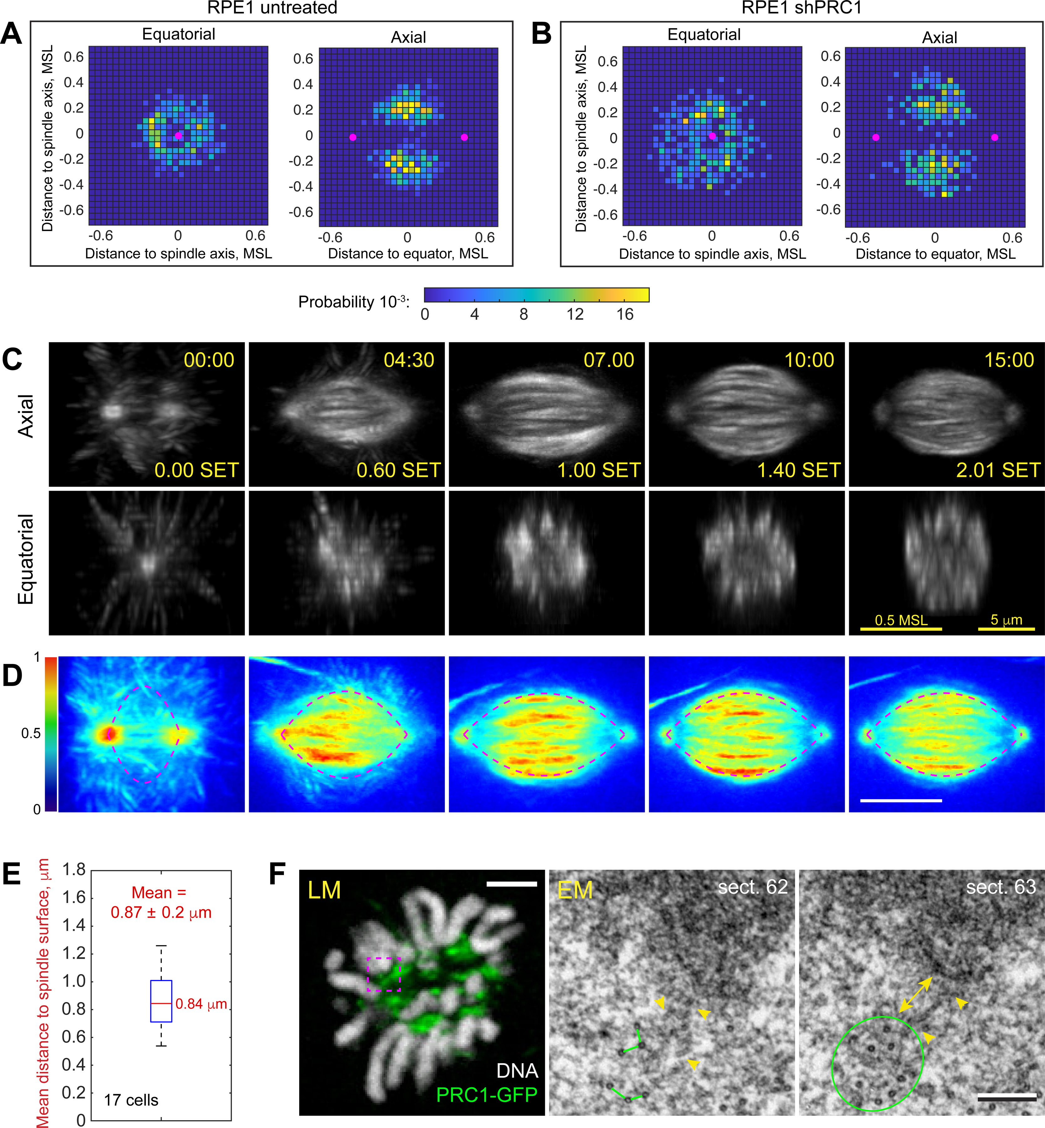
Figure 4. Amphitelic attachments form within a spindle domain enriched in microtubule bundles.

(A,B) Spatial distribution of biorientation events in the untreated (A) and PRC1-depleted (B) cells. 2D histograms in the Equatorial and Axial planes are shown. Distances are normalized to the Maximal Spindle Length in each cell. Magenta dots denote positions of spindle poles at the time with maximum probability of biorientation. (C) Selected timepoints from a timelapse recording of mitosis in RPE1 cells expressing PRC1-GFP. Axial and equatorial maximum-intensity projections of 3D volumes are shown. The volumes are aligned at each timepoint to stabilize the spindle position and orientation. Timestamps are in min:sec after NEB and in fraction of Spindle Elongation Time (SET). Scale bars are 5 μm and 0.5 of the Maximal Spindle Length (MSL) reached in this cell. (D) Average of 3-D time-lapse recordings aligned as in (A) and spatially normalized by the Maximal Spindle Length in each cell. Color map encodes intensity of PRC1-GFP in the averaged volume. Dashed lines approximate the edge of PRC1-enriched domain by an empirically constructed catenary function (see Methods). Timestamps are in SET. Scale bar is 0.5 MSL. (E) Tukey’s boxplot of Euclidian distances from centromeres to the catenary (edge of PRC1-enriched domain) at the time of amphitelic attachment formation. Mean value is reported with STD. (F) Typical arrangement of microtubules near kinetochores adjacent to PRC1-decorated bundles. LM – a single-plane image depicting PRC1-GFP (green) and chromosomes (Hoechst 33342, greyscale) in a fixed cell. EM – 80-nm serial sections through the area boxed in LM. Kinetochore plate is ~250 nm (yellow double arrow) from the edge of a bundle comprising 10 microtubules (green circle) with 50–70-nm spacing between individual microtubules (green lines). Short microtubules (arrowheads) bridge the bundle and the kinetochore plate. See also Video S4.
