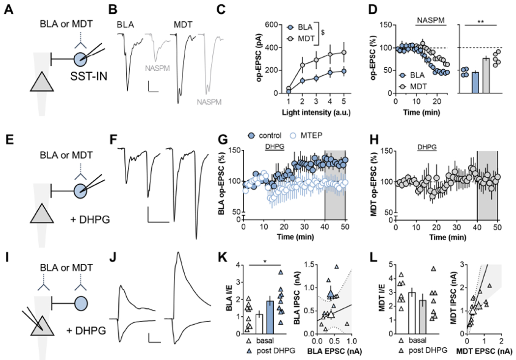
Figure 2. mGlu5 receptor activation enhances amygdalo-cortical feedforward inhibition.

(A) ChR2 was expressed within the basolateral amygdala (BLA) or mediodorsal thalamus (MDT) by viral-mediated gene transfer. Recordings were made from labeled SST-INs and optical (op)-EPSCs were evoked with blue light stimulation. (B) Representative traces of BLA-PFC op-EPSCs at baseline (black) and after NASPM application (gray). Scale bars 50pA, 20ms. (C) MDT synapses onto SST-INs displayed a trend towards larger amplitude op-EPSCs relative to BLA inputs (RM Two-way ANOVA intensity x input interaction: F4,64=2.0, p<0.11; main effect of intensity: F4,64=22.5, p<0.0001; main effect of input: F1,16=3.1, p<0.10). n/N=9/5 cells/mice per group. (D) The Ca2+[ISP CHK]-permeable AMPA receptor antagonist NASPM (200μm, 15min) depressed BLA op-EPSCs onto SST-INs to a greater extent than MDT op-EPSCs (46±4 vs 76±6%; t7=4.2, p<0.004, t-test). n/N = 4-5/4. (E) BLA and MDT op-EPSCs onto SST-INs were elicited before during and after application of the mGlu1/5 agonist DHPG. (F) Representative traces of BLA-PFC op-EPSCs at baseline (left) and after DHPG application (right). Scale bars 100pA, 50ms. (G) The mGlu5 NAM MTEP blocked LTP of BLA-driven op-EPSCs onto SST-INs (107±4 vs 160±9%; t14=5.8, p<0.001, t-test). n/N=5-8/3-5. (H) MDT op-EPSCs did not display LTP (102±15%). n/N=6/3. (I) Feedforward inhibition was assessed in pyramidal cells. (J) Representative traces displaying inward EPSCs evoked at −70 mV and outward inhibitory postsynaptic currents (IPSCs) evoked at 0 mV, at baseline (left) and following DHPG application (right). Scale bars 200pA, 50ms (K) DHPG enhanced the BLA IPSC/EPSC (I/E) ratio in pyramidal cells (1.9 ± 0.3 vs 1.1 ± 0.2; t16=2.16, p<0.05, t-test). n/N=9/3. (L) DHPG had no effect on the MDT-PFC I/E ratio or either current species in isolation n/N = 6/3.
